Orthotic and Prosthetic
Help for Patients & Caregivers
What is Cerebral Palsy?
Cerebral palsy is an umbrella-like term used to describe a group of chronic disorders impairing control of movement that appear in the first few years of life and generally do not worsen over time. The disorders are caused by faulty development of or damage to motor areas in the brain that disrupts the brain’s ability to control movement and posture. Symptoms of cerebral palsy include difficulty with fine motor tasks (such as writing or using scissors), difficulty maintaining balance or walking, involuntary movements. The symptoms differ from person to person and may change over time. Some people with cerebral palsy are also affected by other medical disorders, including seizures or mental impairment, but cerebral palsy does not always cause profound handicap. Early signs of cerebral palsy usually appear before 3 years of age. Infants with cerebral palsy are frequently slow to reach developmental milestones such as learning to roll over, sit, crawl, smile, or walk. Cerebral palsy may be congenital or acquired after birth. Several of the causes of cerebral palsy that have been identified through research are preventable or treatable: head injury, jaundice, Rh incompatibility, and rubella (German measles). Doctors diagnose cerebral palsy by testing motor skills and reflexes, looking into medical history, and employing a variety of specialized tests. Although its symptoms may change over time, cerebral palsy by definition is not progressive, so if a patient shows increased impairment, the problem may be something other than cerebral palsy.
What are the different types of Cerebral Palsy?
Cerebral palsy may be classified by the type of movement problem (such as spastic or athetoid cerebral palsy) or by the body parts involved (hemiplegia, diplegia, and quadriplegia). Spasticity refers to the inability of a muscle to relax, while athetosis refers to an inability to control the movement of a muscle. Infants who at first are hypotonic wherein they are very floppy may later develop spasticity. Hemiplegia is cerebral palsy that involves one arm and one leg on the same side of the body, whereas with diplegia the primary involvement is both legs. Quadriplegia refers to a pattern involving all four extremities as well as trunk and neck muscles. Another frequently used classification is ataxia, which refers to balance and coordination problems. The motor disability of a child with CP varies greatly from one child to another; thus generalizations about children with cerebral palsy can only have meaning within the context of the subgroups described above. For this reason, subgroups will be used in this book whenever treatment and outcome expectations are discussed. Most professionals who care for children with cerebral palsy understand these diagnoses and use them to communicate about a child’s condition.
As noted above, a useful method for making subdivisions is determined by which parts of the body are involved. Although almost all children with cerebral palsy can be classified as having hemiplegia, diplegia, or quadriplegia, there are significant overlaps which have led to the use of additional terms, some of which are very confusing. To avoid confusion, most of the discussion in his book will be limited to the use of these three terms. Occasionally such terms as paraplegia, double hemiplegia, triplegia, and pentaplegia may occasionally be encountered by the reader; these classifications are also based on the parts of the body involved. The dominant type of movement or muscle coordination problem is the other method by which children are subdivided and classified to assist in communicating about the problems of cerebral palsy. The component which seems to be causing the most problem is often used as the categorizing term. For example, the child with spastic diplegia has mostly spastic muscle problems, and most of the involvement is in the legs, but the child may also have a smaller component of athetosis and balance problems. The child with athetoid quadriplegia, on the other hand, would have involvement of both arms and legs, primarily with athetoid muscle problems, but such a child often has some ataxia and spasticity as well. Generally a child with quadriplegia is a child who is not walking independently. The reader may be familiar with other terms used to define specific problems of movement or muscle function terms such as: dystonia, tremor, ballismus, and rigidity. The words severe, moderate, and mild are also often used in combination with both anatomic and motor function classification terms (severe spastic diplegia, for example), but these qualifying words do not have any specific meaning. They are subjective words and their meaning varies depending on the person who is using them.
What causes Cerebral Palsy?
We do not know the cause of most cases of cerebral palsy. That is, we are unable to determine what caused cerebral palsy in most children who have congenital CP. We do know that the child who is at highest risk for developing CP is the premature, very small baby who does not cry in the first five minutes after delivery, who needs to be on a ventilator for over four weeks, and who has bleeding in his brain. Babies who have congenital malformations in systems such as the heart, kidneys, or spine are also more likely to develop CP, probably because they also have malformations in the brain. Seizures in a newborn also increase the risk of CP. There is no combination of factors which always results in an abnormally functioning individual. That is, even the small premature infant has a better than 90 percent chance of not having cerebral palsy. There are a surprising number of babies who have very stormy courses in the newborn period and go on to do very well. In contrast, some infants who have rather benign beginnings are eventually found to have severe mental retardation or learning disabilities.
Is there any treatment?
There is no standard therapy that works for all patients. Drugs can be used to control seizures and muscle spasms, special braces can compensate for muscle imbalance. Surgery, mechanical aids to help overcome impairments, counseling for emotional and psychological needs, and physical, occupational, speech, and behavioral therapy may be employed.
What is the prognosis?
At this time, cerebral palsy cannot be cured, but due to medical research, many patients can enjoy near-normal lives if their neurological problems are properly managed.
For more information on Cerebral Palsy visit:
What is Muscular Dystrophy?
The muscular dystrophies (MD) are a group of genetic diseases characterized by progressive weakness and degeneration of the skeletal muscles that control movement. There are many forms of muscular dystrophy, some noticeable at birth (congenital muscular dystrophy), others in adolescence (Becker MD), but the 3 most common types are Duchenne, facioscapulohumeral, and myotonic. These three types differ in terms of pattern of inheritance, age of onset, rate of progression, and distribution of weakness.
Duchenne MD primarily affects boys and is the result of mutations in the gene that regulates dystrophin – a protein involved in maintaining the integrity of muscle fiber. Onset is between 3-5 years and progresses rapidly. Most boys become unable to walk at 12, and by 20 have to use a respirator to breathe.
Facioscapulohumeral MD appears in adolescence and causes progressive weakness in facial muscles and certain muscles in the arms and legs. It progresses slowly and can vary in symptoms from mild to disabling.
Myotonic MD varies in the age of onset and is characterized by myotonia (prolonged muscle spasm) in the fingers and facial muscles; a floppy-footed, high-stepping gait; cataracts; cardiac abnormalities; and endocrine disturbances. Individuals with myotonic MD have long faces and drooping eyelids; men have frontal baldness.
What are the forms of muscular dystrophy?
The major forms of muscular dystrophy are myotonic, Duchenne, Becker, limb-girdle, facioscapulohumeral, congenital, oculopharyngeal, distal and Emery-Dreifuss.
Some of these names are based on the locations of affected muscles. For example, “facioscapulohumeral” refers to the muscles that move the face, scapula (shoulder blade) and humerus (upper arm bone). Others are based on the type of muscle problem involved (“myotonic” means difficulty relaxing muscles), the age of onset of the disease (as in “congenital,” or birth-onset, dystrophy), or the doctors who first described the disease (Duchenne, Becker, Emery and Dreifuss are doctors’ names).
As the root causes (gene defects) of the muscular dystrophies are discovered, doctors are beginning to change their thinking about how to classify some of the dystrophies. In some cases, a type of muscular dystrophy that looked like it might be one disease has been found to be several different diseases, caused by several different gene defects. This is true for limb-girdle, congenital and distal dystrophies. In other cases, diseases that looked different have been found to be one disease with variations in severity. This is the case with Duchenne and Becker dystrophies.
How do the forms of muscular dystrophy differ?
They differ in severity, age of onset, muscles first and most often affected, the rate at which symptoms progress, and the way the disorders are inherited.
What causes muscular dystrophy?
Flaws in muscle protein genes cause muscular dystrophies. Each cell in our bodies contains tens of thousands of genes. Each gene is a string of the chemical DNA and is the “code” for a protein. (Another way to think of a gene is that it’s the “instructions” or “recipe” for a protein.) If the recipe for a protein is wrong, the protein is made wrong or in the wrong amount or sometimes not at all.
Are muscular dystrophies always inherited?
There is no specific treatment for any of the forms of MD. Respiratory therapy, physical therapy to prevent painful muscle contractures, orthopedic appliances used for support, and corrective orthopedic surgery may be needed to improve the quality of life in some cases. Cardiac abnormalities may require a pacemaker. Corticosteroids such as prednisone can slow the rate of muscle deterioration in patients with Duchenne MD but causes side effects. Myotonia is usually treated with medications such as mexiletine, phenytoin, or quinine.
Is muscular dystrophy contagious?
No. Genetic diseases aren’t contagious.
Is a family medical history important?
Yes. Because the muscular dystrophies can be inherited, it’s important for the doctor to know if anyone in the family ever had a similar disorder.
How is muscular dystrophy diagnosed?
A doctor makes a diagnosis by evaluating the patient’s medical history and by performing a thorough physical examination. Essential to diagnosis are details about when weakness first appeared, its severity, and which muscles are affected. Diagnostic tests may also be used to help the doctor distinguish between different forms of muscular dystrophy, or between muscular dystrophy and other disorders of muscle or nerve.
What are some common diagnostic tests?
Studying a small piece of muscle tissue taken from an individual during a muscle biopsy can sometimes tell a physician whether a disorder is muscular dystrophy and which form of the disease it is.
In Duchenne and Becker muscular dystrophy, a muscle protein called dystrophin is either missing, deficient or abnormally formed. This protein can be examined in the muscle sample.
The reason for the flawed or deficient muscle protein is a flawed gene for dystrophin. A test that involves looking at this gene —DNA testing — can be done to diagnose or rule out Duchenne or Becker muscular dystrophies.
Another diagnostic test is the electromyogram (EMG). To do this test, small electrodes are put into the muscle, which allows the doctor to measure the electrical impulses coming from the muscle. The test is uncomfortable.
Another test often performed measures nerve conduction velocity (NCV). During this test, electrical impulses are sent down the nerves of the arms and legs. By measuring the speed of these impulses with electrodes placed on the skin, the doctor can determine whether the nerves are functioning normally. This test is also uncomfortable.
Blood enzyme tests are helpful because degenerating muscles become “leaky.” They leak enzymes (proteins that speed chemical reactions), which can then be detected in the blood. The presence of these enzymes in the blood at higher than normal levels may be a sign of muscular dystrophy. One such enzyme is creatine kinase, or CK. The CK level is elevated in many forms of muscular dystrophy, some forms resulting in a higher level than others.
Is there any treatment?
There is no specific treatment for any of the forms of MD. Respiratory therapy, physical therapy to prevent painful muscle contractures, orthopedic appliances used for support, and corrective orthopedic surgery may be needed to improve the quality of life in some cases. Cardiac abnormalities may require a pacemaker. Corticosteroids such as prednisone can slow the rate of muscle deterioration in patients with Duchenne MD but causes side effects. Myotonia is usually treated with medications such as mexiletine, phenytoin, or quinine.
For more information on Muscular Dystrophy visit:
What is Plagiocephaly?
Plagiocephaly is a “malformation of the head marked by an oblique slant to the main axis of the skull”. The Term has also been applied to any condition characterized by a persistent flatten spot on the back or one side of the child’s head.
What causes Plagiocephaly?
Up until about one year of age, the bones of your baby’s head are very thin and flexible. This makes your baby’s head very soft and easy to mold. For the first few months of life your baby will not be strong enough to roll over on his own. If your baby prefers to look in one direction or if your baby is always on his back, part of his skull may become flat. This flattening is caused by constant pressure on one part of the skull. This is called positional plagiocephaly. Your baby may also develop a flat spot if he spends long periods of time in a car seat or reclining seat.
You may start to see flattening when your baby is only four to six weeks old.
What is the diagnosis?
The diagnosis begins with an examination by a pediatrician, pediatric neurosurgeon or craniofacial surgeon. A primary objective of the examination is to rule out craniosynostosis (a condition that requires surgical correction). The initial examination involves questions about gestation and birth, in utero position, neck tightness and post-natal positioning (for example, sleeping position). The physical examination includes inspection of the infant’s head and may involve palpation (carefully feeling) of the child’s skull for suture ridges and soft spots (the fontanelles) as well as checking for neck tightness and other deformities. The physician may also request x-rays or computerized tomography (a CAT scan, a series of photographic images of the skull). These images provide the most reliable method for diagnosing premature suture fusion (craniosynostosis). In addition, the physician may make (or order) a series of measurements from the child’s face and head [more on cranial anthropometry]. These measurements will be used to assess severity and monitor treatment.
Is there any treatment?
To prevent your baby from developing a flattened skull, change his position often. Put your baby on his tummy to play several times a day. Use a firm play surface such as a carpeted floor or an activity mat on the floor. “Tummy time” will also help your baby:
Develop early control of his head
Strengthen the muscles in the upper body
Learn to roll over
Reach for objects
Learn to crawl
You can also put your baby on his side to play. To keep your baby on his side, put a firm rolled-up towel or blanket behind his back.
Treatment for deformational plagiocephaly
Specific treatment will be determined by your child’s physician based on the severity of the deformational plagiocephaly. Frequent rotation of your child’s head would be the first recommendation once your infant has been diagnosed with plagiocephaly. Alternating your infant’s sleep position from the back to the sides, and not putting infants on their backs when they are awake may also help prevent and treat positional plagiocephaly. Some cases do not require any treatment and the condition may resolve spontaneously when the infant begins to sit.
If the deformity is moderate to severe and a trial of re-positioning has failed, your child’s physician may recommend a cranial remodeling band or helmet.
How does helmeting correct deformational plagiocephaly?
Helmets are usually made of an outer hard shell with a foam lining. Gentle, persistent pressures are applied to capture the natural growth of an infant’s head, while inhibiting growth in the prominent areas and allowing for growth in the flat regions. As the head grows, adjustments are made frequently. The helmet essentially provides a tight, round space for the head to grow into.
How long will my child wear a helmet?
The average treatment with a helmet is usually three to six months, depending on the age of the infant and the severity of the condition. Careful and frequent monitoring is required. Helmets must be prescribed by a licensed physician with craniofacial experience.
For more information on Plagiocephaly visit:
Lucile Packard Children’s Hospital at Stanford
Plagiocephaly.info
What is Scoliosis?
Scoliosis is not a disease – it is a descriptive term. All spines have curves. Some curvature in the neck, upper trunk and lower trunk is normal. Humans need these spinal curves to help the upper body maintain proper balance and alignment over the pelvis. However, when there are abnormal side-to-side (lateral) curves in the spinal column, we refer to this as scoliosis.
What to look for?
There are several different “warning signs” to look for to help determine if you or someone you love has scoliosis. Should you notice any one or more of these signs, you should schedule an exam with a doctor.
Shoulders are different heights – one shoulder blade is more prominent than the other
Head is not centered directly above the pelvis
Appearance of a raised, prominent hip
Rib cages are at different heights
Uneven waist
Changes in look or texture of skin overlying the spine (dimples, hairy patches, color changes)
Leaning of entire body to one side
What causes Scoliosis?
Doctors define scoliosis in a particular person based on a number of factors related to the curve, including:
Shape. Aside from appearing like the letter C or S, a curve may occur in two or three dimensions. A nonstructural curve is a side-to-side curve. A structural curve involves twisting of the spine and occurs in three dimensions.
Location. The curve may occur in the upper back area (thoracic), the lower back area (lumbar) or in both areas (thoracolumbar).
Direction. The curve can bend to the left or to the right.
Angle. Doctors figure out the angle of the curve using the vertebra at the apex of the curve as the starting point.
Cause. About 80 percent of scoliosis cases are idiopathic, meaning the cause is unknown.
Many theories have been proposed regarding the causes of scoliosis. They include connective tissue disorders, hormonal imbalance and abnormality in the nervous system.
Scoliosis runs in families and may involve genetic (hereditary) factors. But researchers haven’t identified the gene or genes that may cause scoliosis. Doctors also recognize that spinal cord and brainstem abnormalities play a role in some cases of scoliosis.
Is there any treatment?
There are three basic types of treatments for scoliosis: observation, orthopedic bracing, or surgery.
Observation is appropriate for small curves, curves that are at low risk of progression, and those with a natural history that is favorable at the completion of growth. These decisions are based on the expected natural history of a given curve. For example, if your child is diagnosed with a curve of 25 to 40 degrees and has completed growth (i.e., boys older than 17, girls older than 15), then observation is appropriate. Statistically, these curves are at low risk of progression and are not likely to cause problems in adulthood. Follow-up x-ray once per year for several years would then confirm that the curve is not progressing after completion of growth. As an adult, an x-ray every five years, or if there are symptoms, is sufficient.
Orthopedic braces are used to prevent further spinal deformity in children with curve magnitudes within the range of 25 to 40 degrees. If these children already have curvatures of these magnitudes and still have a substantial amount of skeletal growth left, then bracing is a viable option. It is important to note, however, that the intent of bracing is to prevent further deformity – it is not to correct the existing curvature or to make the curve disappear.
Surgery is an option used primarily for severe scoliosis (curves greater than 45 degrees) or for curves that do not respond to bracing. There are two primary goals for surgery: to stop a curve from progressing during adult life and to diminish spinal deformity.
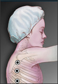
Until the last few decades, patients undergoing scoliosis surgery endured intensive surgery, treatment and casting, as well as months of slow recuperation. Since that time, spinal surgery pioneers such as Paul Harrington, Yves Paul Cotrel and Jean Dubousset have made great strides in improving the techniques and instruments used in surgery and post-operative care for patients with scoliosis.
There are different techniques and methods used today for scoliosis surgery. The most frequently performed surgery for adolescent idiopathic scoliosis involves posterior spinal fusion with instrumentation and bone grafting. This kind of surgery is performed through the patient’s back while the patient lies on his or her stomach. Two common instrumentation techniques are called Cotrel-Dubousset (CD®) instrumentation (rod rotation technique) and COLORADO™ instrumentation (translation technique). During these types of surgery, the surgeon attaches a metal rod to each side of the patient’s spine by using hooks attached to the vertebral bodies. Then, the surgeon fuses the spine with a piece of bone from the patient’s hip (a bone graft). The bone grows in between the vertebrae and holds them together and straight. This process is called spinal fusion. The metal rods attached to the spine ensure that the backbone remains straight while the spinal fusion takes place.
The operation usually takes several hours. With recent advances in technology, most people with idiopathic scoliosis are released within a week of surgery and do not require post-operative bracing. Most patients are able to return to school or work in two to four weeks after the surgery and are able to resume all pre-operative activities within four to six months.
Another surgery option for scoliosis is an anterior approach, which means that the surgery is conducted through the chest walls instead of entering through the patient’s back. The patient lies on his or her side during the surgery. During this procedure, the surgeon makes incisions in the patient’s side, deflates the lung and removes a rib in order to reach the spine. This approach allows the surgeon to operate higher up in the spine than through posterior approaches, and studies have shown favorable results with this type of surgery. Video-assisted thoracoscopic surgery allows surgeons to enhance their vision of the spine and to conduct a less invasive surgery than with an open procedure. The anterior spinal approach has several advantages: better cosmetic results, quicker patient rehabilitation, improved spine mobilization, and fusion of fewer segments. Most patients require bracing for several months after this surgery.
What is the prognosis?
At this time, cerebral palsy cannot be cured, but due to medical research, many patients can enjoy near-normal lives if their neurological problems are properly managed.
For more information on Scoliosis visit:
What is Spinal Cord Injury?
Spinal Cord Injury (SCI) is damage to the spinal cord that results in a loss of function such as mobility or feeling. Frequent causes of damage are trauma (car accident, gunshot, falls, etc.) or disease (polio, spina bifida, Friedreich’s Ataxia, etc.). The spinal cord does not have to be severed in order for a loss of functioning to occur. In fact, in most people with SCI, the spinal cord is intact, but the damage to it results in loss of functioning. SCI is very different from back injuries such as ruptured disks, spinal stenosis or pinched nerves.
A person can “break their back or neck” yet not sustain a spinal cord injury if only the bones around the spinal cord (the vertebrae) are damaged, but the spinal cord is not affected. In these situations, the individual may not experience paralysis after the bones are stabilized.
What is the Spinal Cord and the Vertebra?
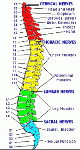
The spinal cord is about 18 inches long and extends from the base of the brain, down the middle of the back, to about the waist. The nerves that lie within the spinal cord are upper motor neurons (UMNs) and their function is to carry the messages back and forth from the brain to the spinal nerves along the spinal tract. The spinal nerves that branch out from the spinal cord to the other parts of the body are called lower motor neurons (LMNs). These spinal nerves exit and enter at each vertebral level and communicate with specific areas of the body. The sensory portions of the LMN carry messages about sensation from the skin and other body parts and organs to the brain. The motor portions of the LMN send messages from the brain to the various body parts to initiate actions such as muscle movement.
The spinal cord is the major bundle of nerves that carry nerve impulses to and from the brain to the rest of the body. The brain and the spinal cord constitute the Central Nervous System. Motor and sensory nerves outside the central nervous system constitute the Peripheral Nervous System, and another diffuse system of nerves that control involuntary functions such as blood pressure and temperature regulation are the Sympathetic and Parasympathetic Nervous Systems.
The spinal cord is surrounded by rings of bone called vertebra. These bones constitute the spinal column (back bones). In general, the higher in the spinal column the injury occurs, the more dysfunction a person will experience. The vertebra are named according to their location. The eight vertebra in the neck are called the Cervical Vertebra. The top vertebra is called C-1, the next is C-2, etc. Cervical SCI’s usually cause loss of function in the arms and legs, resulting in quadriplegia. The twelve vertebra in the chest are called the Thoracic Vertebra. The first thoracic vertebra, T-1, is the vertebra where the top rib attaches.
Injuries in the thoracic region usually affect the chest and the legs and result in paraplegia. The vertebra in the lower back between the thoracic vertebra, where the ribs attach, and the pelvis (hip bone), are the Lumbar Vertebra. The sacral vertebra run from the Pelvis to the end of the spinal column. Injuries to the five Lumbar vertebra (L-1 thru L-5) and similarly to the five Sacral Vertebra (S-1 thru S-5) generally result in some loss of functioning in the hips and legs.
What are the effects of SCI? The effects of SCI depend on the type of injury and the level of the injury. SCI can be divided into two types of injury – complete and incomplete. A complete injury means that there is no function below the level of the injury; no sensation and no voluntary movement. Both sides of the body are equally affected. An incomplete injury means that there is some functioning below the primary level of the injury. A person with an incomplete injury may be able to move one limb more than another, may be able to feel parts of the body that cannot be moved, or may have more functioning on one side of the body than the other. With the advances in acute treatment of SCI, incomplete injuries are becoming more common.
The level of injury is very helpful in predicting what parts of the body might be affected by paralysis and loss of function. Remember that in incomplete injuries there will be some variation in these prognoses.
Cervical (neck) injuries usually result in quadriplegia. Injuries above the C-4 level may require a ventilator for the person to breathe. C-5 injuries often result in shoulder and biceps control, but no control at the wrist or hand. C-6 injuries generally yield wrist control, but no hand function. Individuals with C-7 and T-1 injuries can straighten their arms but still may have dexterity problems with the hand and fingers. Injuries at the thoracic level and below result in paraplegia, with the hands not affected. At T-1 to T-8 there is most often control of the hands, but poor trunk control as the result of lack of abdominal muscle control. Lower T-injuries (T-9 to T-12) allow good truck control and good abdominal muscle control. Sitting balance is very good. Lumbar and Sacral injuries yield decreasing control of the hip flexors and legs.
Besides a loss of sensation or motor functioning, individuals with SCI also experience other changes. For example, they may experience dysfunction of the bowel and bladder,. Sexual functioning is frequently with SCI may have their fertility affected, while women’s fertility is generally not affected. Very high injuries (C-1, C-2) can result in a loss of many involuntary functions including the ability to breathe, necessitating breathing aids such as mechanical ventilators or diaphragmatic pacemakers. Other effects of SCI may include low blood pressure, inability to regulate blood pressure effectively, reduced control of body temperature, inability to sweat below the level of injury, and chronic pain.
What is Spinal Stenosis?
The spinal canal is like a tunnel which runs up and down the human spine. This canal sits directly behind the bony blocks which make up the spine (vertebrae) and contains the nerves (spinal cord and nerve roots) running from the brain to all areas of the body
When something causes a narrowing of this canal then the nerves can become irritated or squeezed. This can lead to a variety of symptoms ranging from tingling, numbness, and weakness to severe pain and paralysis. Common conditions which can narrow the spinal canal include a herniated disc (often called a slipped disc), fracture of the spine, tumor, infection and degeneration. A set of symptoms related to narrowing of the spinal canal seen with aging and degeneration is called spinal stenosis. The symptoms of spinal stenosis most commonly include a sensation of heaviness, weakness and pain with walking or prolonged standing. At rest these symptoms usually disappear. These symptoms are related to the irritation of the nerves in the spinal canal which is worsened with standing or walking due to mechanical compression or stretching of the nerves. Patients often complain of a gradual decrease in their ability to walk, requiring more frequent stops to rest their legs. The treatment for spinal stenosis is dependant on the severity of symptoms. Generally, aerobic activities like walking combined with a guided exercise program and weight loss (in overweight patients) is recommended first.
When there is no relief, some specialists recommend injection treatments although the effectiveness of this is limited. Surgery is indicated when symptoms are severe, progressive and a specific area of narrowing in the spinal canal has been discovered. The surgical procedure is aimed at freeing up the nerves in the canal by removing pieces of bone and thickened tissues such as the ligaments. A spinal fusion may also be necessary to stabilize the spine
The spine consists of a series of bone blocks (vertebral bodies) which are separated from one another by discs of soft tissue. Within the structure of the spine sits a tunnel called the spinal canal. This tunnel contains the neurologic structures including the spinal cord and nerve roots. Although there is some free space between the neurologic structures and the edges of the spinal canal, this space can be reduced by many different conditions including injury to the spine. The canal is surrounded by bone and ligaments and therefore can not expand if the spinal cord or nerves require more room. Therefore, if anything begins to narrow the spinal canal, there is risk for irritation or injury of the spinal cord or nerves. Conditions which can lead to narrowing of the spinal canal include infection, tumors, trauma, herniated disc, arthritis and degeneration.
Spinal stenosis refers to the condition of neurologic problems associated with narrowing of the spinal canal due to degenerative changes in the spine. Arthritis of the small joints in the spine (facets) as well as thickening of ligaments and formation of bony spurs can all lead to gradual squeezing and irritation of neurologic structures. This process is usually gradual and can lead to symptoms such as pain with walking, a decreased endurance for physical activities, heaviness in the legs, tingling sensations, tightness and numbness in the legs with activity, and often associated low back pains.
Treatment for spinal stenosis ranges from physical therapy to epidural injections and finally surgery in certain cases. Since patients affected by spinal stenosis are usually elderly, treatment must carefully consider not only the disease in the spine but also the risks and benefits of treatment in each individual. Although therapy and steroid injections into the affected area of the spine can offer good relief in some patients, there are people who will only get temporary relief if at all. In patients who have failed non-operative treatment, surgery can sometimes be considered. Prior to designing a treatment plan for any individual, careful diagnosis must be made. This will often involve tests such as an MRI, CT scan, or myelogram and plain X-rays. In those patients who are candidates for surgery, the goal is to free up the constricted regions of the spinal canal to ensure freeing the affected neurologic structures. Occasionally, in order to stabilize a degenerated part of the spine, a fusion will be performed. This involves laying down of bone over an area of the spine so that a solid block is created where there was previously arthritis with pain and an unstable spine.
Surgery for spinal stenosis has a high success rate in patients carefully selected for this procedure. It remains a useful approach in treatment when other options have been exhausted and after careful review of risks and benefits with the patient.
What are the effects of SCI?
The effects of SCI depend on the type of injury and the level of the injury. SCI can be divided into two types of injury – complete and incomplete. A complete injury means that there is no function below the level of the injury; no sensation and no voluntary movement. Both sides of the body are equally affected. An incomplete injury means that there is some functioning below the primary level of the injury. A person with an incomplete injury may be able to move one limb more than another, may be able to feel parts of the body that cannot be moved, or may have more functioning on one side of the body than the other. With the advances in acute treatment of SCI, incomplete injuries are becoming more common.
The level of injury is very helpful in predicting what parts of the body might be affected by paralysis and loss of function. Remember that in incomplete injuries there will be some variation in these prognoses.
Cervical (neck) injuries usually result in quadriplegia. Injuries above the C-4 level may require a ventilator for the person to breathe. C-5 injuries often result in shoulder and biceps control, but no control at the wrist or hand. C-6 injuries generally yield wrist control, but no hand function. Individuals with C-7 and T-1 injuries can straighten their arms but still may have dexterity problems with the hand and fingers. Injuries at the thoracic level and below result in paraplegia, with the hands not affected. At T-1 to T-8 there is most often control of the hands, but poor trunk control as the result of lack of abdominal muscle control. Lower T-injuries (T-9 to T-12) allow good truck control and good abdominal muscle control. Sitting balance is very good. Lumbar and Sacral injuries yield decreasing control of the hip flexors and legs.
Besides a loss of sensation or motor functioning, individuals with SCI also experience other changes. For example, they may experience dysfunction of the bowel and bladder,. Sexual functioning is frequently with SCI may have their fertility affected, while women’s fertility is generally not affected. Very high injuries (C-1, C-2) can result in a loss of many involuntary functions including the ability to breathe, necessitating breathing aids such as mechanical ventilators or diaphragmatic pacemakers. Other effects of SCI may include low blood pressure, inability to regulate blood pressure effectively, reduced control of body temperature, inability to sweat below the level of injury, and chronic pain.
How many people have SCI?
Approximately 450,000 people live with SCI in the US. There are about 10,000 new SCI’s every year; the majority of them (82%) involve males between the ages of 16-30. These injuries result from motor vehicle accidents (36%), violence (28.9%), or falls (21.2%).Quadriplegia is slightly more common than paraplegia.
For more information on Spinal Cord Injury visit:
What is Vascular Disease?
The vascular system is the network of blood vessels that circulate blood to and from the heart and lungs. Vascular diseases are very common, especially as people age. Many people have these diseases and don’t know it, because they rarely cause symptoms in the early stages. People with risk factors or any signs or symptoms of vascular disease, should be evaluated by a physician. Untreated vascular disease can lead to serious health problems, such as tissue death and gangrene requiring amputation or other surgery; chronic disability and pain; and weakened blood vessels that may rupture without warning. Deadly complications can result, including stroke (a clogged or narrowed blood vessel cuts the supply of blood to the brain), and pulmonary embolism (a blood clot breaks loose and travels to the heart and lungs).
What are the symptoms of Vascular Disease?
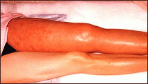
Swelling and discoloration of the leg is a sign of Deep Vein Thrombosis
There may be no symptoms of venous disease caused by blood clots until the clot grows large enough to block the flow of blood through the vein. Symptoms may then come on suddenly:
Pain
Sudden swelling in the affected limb
Enlargement of the superficial veins
Reddish-blue discoloration
Skin that is warm to the touch
What are the symptoms of Pulmonary Embolism?
A sudden feeling of apprehension
Shortness of breath
Sharp chest pain
Rapid pulse
Sweating
Cough with bloody sputum
Fainting
If you experience the sudden onset of any of these symptoms, contact your doctor or seek emergency treatment immediately!
Varicose veins , also called “varicoceles,” result when the valves that control the flow of blood in and out of veins fail to work properly and the pull of gravity causes blood to pool in the legs or elsewhere. Varicoceles in the scrotum may cause infertility in men. Varicoceles in the veins of the ovaries may cause chronic pelvic pain in some women.
When valves fail in the legs, the superficial veins become enlarged and twisted, where they appear as twisted, dark blue vessels just under the skin’s surface. Smaller varicose veins are sometimes called spider veins. Obesity, pregnancy, constriction of the veins with garters or tight clothing, and an inherited tendency are among the contributing causes of varicose veins. Usually, there are no symptoms. Varicose veins are diagnosed by physical examination.
Women between the ages of 30 and 70 are most often affected by Varicose Veins. In the United States, 10 percent of men and 20 percent of women have varicose or spider veins. Treatment usually is not required. While most treatment is sought for cosmetic reasons – to improve the appearance of the veins in the legs – some varicose veins are painful and require treatment for medical reasons.
Symptoms of Varicose Veins
Most varicose veins have no symptoms other than the appearance of purplish, knotted veins on the surface of the skin. A physician should be consulted and treatment may be required if there is:
Pain or heaviness in the leg, feet and ankles,
Swelling,
Sores or ulcers on the skin, or
Severe bleeding if the vein is injured.
Phlebitis is an inflammation of a vein that can be due to bacterial infection, injury or unknown causes. Thrombophlebitis is inflammation that results from the formation of a blood clot in an arm or leg vein. It can occur in a superficial vein near the skin surface or in a deep vein. Pain and inflammation are the most common symptoms. Unfortunately, in the case of thrombophlebitis in the deep veins (see deep vein thrombosis) there may be no symptoms unless the clot travels to the lungs, resulting in a life-threatening pulmonary embolism.
Venous stasis disease also is caused by defective values in the veins, but it is far more serious than varicose veins. If a damaged valve does not close completely, pooled blood can build up in the veins causing pain, swelling and tissue damage that may lead to painful sores or ulcers. Chronic venous stasis disease can result in devastating disfigurement, disability and a lifetime of treatments and hospital stays. Fortunately, early diagnosis and treatment can avoid these long-term effects.
Diagnosing Vascular Disease?
Diagnosing Venous Disease and Pulmonary Embolism
Venous disease is diagnosed using one or more of the following techniques:
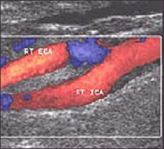
Ultrasound is a technique in which a “transducer” (a hand held device about the size of a computer mouse) is moved over the skin and harmless sound waves “bounce” back signals that are computerized to create an image. The technique is painless and has no known risk. Here, a “colorized” ultrasound image highlights the blood vessels.
Duplex or Doppler Ultrasound – This non-invasive technique uses ultrasound to “see” clots or other abnormalities in the blood vessels.
CT Scan (Computed Tomography) is similar to an X-ray except the images are computerized to appear as a series of slides. When viewed together, the slices provide a three-dimensional image. Sometimes a special dye, or contrast agent, is injected or swallowed before the exam to highlight the images.
Venography is a type of X-ray (called angiography) in which a thin, flexible tube, or catheter, is threaded into the blood vessels. A local anesthetic is given to numb the skin where the catheter is inserted, and X-rays are used to guide the catheter. A contrast agent, or dye, is injected through the catheter to highlight the blood vessel and call attention to any abnormalities. This procedure is performed by an interventional radiologist – a specialist who diagnoses and treats many vascular diseases and other conditions without surgery.
Magnetic Resonance Angiography (MRA) is a noninvasive exam in which a magnetic resonance (MR) scanner uses harmless but powerful magnetic fields and radio waves to create detailed images of the blood vessels.
Diagnosing Pulmonary Embolism
V/Q Scan (sometimes called a V/P or ventilation/perfusion scan) is a nuclear medicine test in which short-acting radioactive particles are injected through a vein or breathed into the lungs. If there are areas of the lung that do not “take up” the particles, it is an indication that there may be a blood clot. Computed tomography (CT), chest X-rays or venography also may be used to diagnose blood clots in the lung.
What Causes Vascular Disease?
Risk Factors that increase the chances of venous disease include:
A family history.
Increasing age that results in a loss of elasticity in the veins and their valves
Pregnancy
Illness or injury
Prolonged periods of inactivity – sitting, standing or bed rest.
Hypertension, diabetes, high cholesterol
Other conditions that affect the health of the cardiovascular system
Smoking
Obesity
Preventing Vascular Disease
The best way to prevent vascular disease is to live a “heart healthy” lifestyle – don’t smoke; eat nutritious, low fat foods; exercise; control risk factors and maintain a healthy weight.
Life style changes. The single most effective steps you can take to prevent vascular disease are to quit smoking and control high blood pressure, high cholesterol, diabetes and other factors that contribute to vascular disease. Regular exercise, eating a balanced diet and maintaining a healthy weight also are important.
For more information on Vascular Disease visit:
What is type 2 diabetes?
Diabetes is a disease in which blood glucose levels are above normal. People with diabetes have problems converting food to energy. After a meal, food is broken down into a sugar called glucose, which is carried by the blood to cells throughout the body. Cells use the hormone insulin, made in the pancreas, to help them process blood glucose into energy.
People develop type 2 diabetes because the cells in the muscles, liver, and fat do not use insulin properly. Eventually, the pancreas cannot make enough insulin for the body’s needs. As a result, the amount of glucose in the blood increases while the cells are starved of energy. Over the years, high blood glucose damages nerves and blood vessels, leading to complications such as heart disease, stroke, blindness, kidney disease, nerve problems, gum infections, and amputation.
How can type 2 diabetes be prevented?
Although people with diabetes can prevent or delay complications by keeping blood glucose levels close to normal, preventing or delaying the development of type 2 diabetes in the first place is even better. The results of a major federally funded study, the Diabetes Prevention Program (DPP), show how to do so. This study of 3,234 people at high risk for diabetes showed that moderate diet and exercise resulting in a 5- to 7-percent weight loss can delay and possibly prevent type 2 diabetes.
Study participants were overweight and had higher than normal levels of blood glucose, a condition called pre-diabetes (impaired glucose tolerance). Both pre-diabetes and obesity are strong risk factors for type 2 diabetes.
Am I at Risk for Type 2 Diabetes?
Because of the high risk among some minority groups, about half of the DPP participants were African American, American Indian, Asian American, Pacific Islander, or Hispanic American/Latino. The DPP tested two approaches to preventing diabetes: a healthy eating and exercise program (lifestyle changes), and the diabetes drug metformin. People in the lifestyle modification group exercised about 30 minutes a day 5 days a week (usually by walking) and lowered their intake of fat and calories. Those who took the diabetes drug metformin received standard information on exercise and diet. A third group received only standard information on exercise and diet.
The results showed that people in the lifestyle modification group reduced their risk of getting type 2 diabetes by 58 percent. Average weight loss in the first year of the study was 15 pounds. Lifestyle modification was even more effective in those 60 and older. They reduced their risk by 71 percent. People receiving metformin reduced their risk by 31 percent.
What are the signs and symptoms of type 2 diabetes?
Many people have no signs or symptoms. Symptoms can also be so mild that you might not even notice them. Nearly six million people in the United States have type 2 diabetes and do not know it.
Here is what to look for:
increased thirst
increased hunger
fatigue
increased urination, especially at night
weight loss
blurred vision
sores that do not heal
Types of Diabetes
The three main kinds of diabetes are type 1, type 2, and gestational diabetes.
Type 1 Diabetes
Type 1 diabetes, formerly called juvenile diabetes or insulin-dependent diabetes, is usually first diagnosed in children, teenagers, or young adults. In this form of diabetes, the beta cells of the pancreas no longer make insulin because the body’s immune system has attacked and destroyed them. Treatment for type 1 diabetes includes taking insulin shots or using an insulin pump, making wise food choices, exercising regularly, taking aspirin daily (for some), and controlling blood pressure and cholesterol.
Type 2 Diabetes
Type 2 diabetes, formerly called adult-onset or noninsulin-dependent diabetes, is the most common form of diabetes. People can develop type 2 diabetes at any age, even during childhood. This form of diabetes usually begins with insulin resistance, a condition in which fat, muscle, and liver cells do not use insulin properly. At first, the pancreas keeps up with the added demand by producing more insulin. In time, however, it loses the ability to secrete enough insulin in response to meals. Being overweight and inactive increases the chances of developing type 2 diabetes. Treatment includes taking diabetes medicines, making wise food choices, exercising regularly, taking aspirin daily, and controlling blood pressure and cholesterol.
Gestational Diabetes
Some women develop gestational diabetes during the late stages of pregnancy. Although this form of diabetes usually goes away after the baby is born, a woman who has had it is more likely to develop type 2 diabetes later in life. Gestational diabetes is caused by the hormones of pregnancy or a shortage of insulin.
Am I at Risk for Type 2 Diabetes?
Sometimes people have symptoms but do not suspect diabetes. They delay scheduling a checkup because they do not feel sick. Many people do not find out they have the disease until they have diabetes complications, such as blurry vision or heart trouble. It is important to find out early if you have diabetes because treatment can prevent damage to the body from diabetes.
Should I be tested for diabetes?
Anyone 45 years old or older should consider getting tested for diabetes. If you are 45 or older and overweight (see BMI chart on pages 10 and 11), it is strongly recommended that you get tested. If you are younger than 45, overweight, and have one or more of the risk factors on page 5, you should consider testing. Ask your doctor for a fasting blood glucose test or an oral glucose tolerance test. Your doctor will tell you if you have normal blood glucose, pre-diabetes, or diabetes.
What does it mean to have pre-diabetes?
It means you are at risk for getting type 2 diabetes and heart disease. The good news is if you have pre-diabetes you can reduce the risk of getting diabetes and even return to normal blood glucose levels. With modest weight loss and moderate physical activity, you can delay or prevent type 2 diabetes. If your blood glucose is higher than normal but lower than the diabetes range (what we now call pre-diabetes), have your blood glucose checked in 1 to 2 years.
Doing My Part: Getting Started
Making big changes in your life is hard, especially if you are faced with more than one change. You can make it easier by taking these steps:
Make a plan to change behavior.
Decide exactly what you will do and when you will do it.
Plan what you need to get ready.
Think about what might prevent you from reaching your
goals.Find family and friends who will support and encourage you.
Decide how you will reward yourself when you do what you
have planned.
Your doctor, a dietitian, or a counselor can help you make a plan. Here are some of the areas you may wish to change to reduce your risk of diabetes.
Reach and Maintain a Reasonable Body Weight
Your weight affects your health in many ways. Being overweight can keep your body from making and using insulin properly. It can also cause high blood pressure. The DPP showed that losing even a few pounds can help reduce your risk of developing type 2 diabetes because it helps your body use insulin more effectively. In the DPP, people who lost between 5 and 7 percent of their body weight significantly reduced their risk of type 2 diabetes. For example, if you weigh 200 pounds, losing only 10 pounds could make a difference.
Body mass index (BMI) is a measure of body weight relative to height. You can use BMI to see whether you are underweight, normal weight, overweight, or obese. Click here to view the BMI chart.
Find your height in the left-hand column.
Move across in the same row to the number closest to your weight.
The number at the top of that column is your BMI. Check the word above your BMI to see whether you are normal weight, overweight, or obese.
If you are overweight or obese, choose sensible ways to get in shape:
Avoid crash diets. Instead, eat less of the foods you usually have. Limit the amount of fat you eat.
Increase your physical activity. Aim for at least 30 minutes of exercise most days of the week.
Set a reasonable weight-loss goal, such as losing 1 pound a week. Aim for a long-term goal of losing 5 to 7 percent of your total body weight.
Make Wise Food Choices Most of the Time
What you eat has a big impact on your health. By making wise food choices, you can help control your body weight, blood pressure, and cholesterol.
Take a hard look at the serving sizes of the foods you eat. Reduce serving sizes of main courses (such as meat), desserts, and foods high in fat. Increase the amount of fruits and vegetables.
Limit your fat intake to about 25 percent of your total calories. For example, if your food choices add up to about 2,000 calories a day, try to eat no more than 56 grams of fat. Your doctor or a dietitian can help you figure out how much fat to have. You can check food labels for fat content too.
You may also wish to reduce the number of calories you have each day. People in the DPP lifestyle modification group lowered their daily calorie total by an average of about 450 calories. Your doctor or dietitian can help you with a meal plan that emphasizes weight loss.
Keep a food and exercise log. Write down what you eat, how much you exercise—anything that helps keep you on track.
When you meet your goal, reward yourself with a nonfood item or activity, like watching a movie.
Be Physically Active Every Day
Regular exercise tackles several risk factors at once. It helps you lose weight, keeps your cholesterol and blood pressure under control, and helps your body use insulin. People in the DPP who were physically active for 30 minutes a day 5 days a week reduced their risk of type 2 diabetes. Many chose brisk walking for exercise.
If you are not very active, you should start slowly, talking with your doctor first about what kinds of exercise would be safe for you. Make a plan to increase your activity level toward the goal of being active at least 30 minutes a day most days of the week. Choose activities you enjoy. Here are some ways to work extra activity into your daily routine:
Take the stairs rather than an elevator or escalator.
Park at the far end of the lot and walk.
Get off the bus a few stops early and walk the rest of the way.
Walk or bicycle instead of drive whenever you can.
Take Your Prescribed Medications
Some people need medication to help control their blood pressure or cholesterol levels. If you do, take your medicines as directed. Ask your doctor whether there are any medicines you can take to prevent type 2 diabetes.
Hope Through Research
We now know that many people can prevent type 2 diabetes through weight loss, regular exercise, and lowering their intake of fat and calories. Researchers are intensively studying the genetic and environmental factors that underlie the susceptibility to obesity, pre-diabetes, and diabetes. As they learn more about the molecular events that lead to diabetes, they will develop ways to prevent and cure the different stages of this disease. People with diabetes and those at risk for it now have easier access to clinical trials that test promising new approaches to treatment and prevention. For information about current studies, see ClinicalTrials.gov.
Diabetes – Staying healthy from head to toe
If you have diabetes, controlling your sugar is always the first priority. A healthy diet, regular exercise and good medical care can help. When your blood sugar is under control you’re also at lower risk for complications from diabetes. High blood sugar levels can damage your nerves and blood vessels. When levels are too high it can cause damage and disease in your eyes, teeth and feet. That’s why these parts of your body need special care, according to the American Diabetes Association.
Eyes. To keep your eyes healthy, get an eye exam every year. You should also go to the doctor if:
Your vision gets blurry.
You see double.
Your eyes hurt.
You see spots.
Teeth and gums. Have your teeth cleaned and checked every 6 months. Brush your teeth, front and back, twice daily with a soft brush. Floss once a day. See your dentist if you notice any problems with your gums or teeth.
Feet. Wash and dry your feet every day. Use lotion to keep the skin from drying out. Check every day for sores, blisters, calluses or swelling. Don’t try to treat calluses or corns at home. See your doctor. Cut toenails straight across. Look for sharp edges—they can cut your Check shoes inside and out for sharp objects before you put them on. Pebbles, nails or even a torn shoe lining could cause problems.